
Just as we were about to celebrate Chinese New Year, the last case presented with an arrhythmia, which is uncommonly seen in our practice.
She is a 14-year-old Labrador, referred for further evaluation due to exercise intolerance and detection of an abnormal heart rhythm.
Summary of Findings
Blood Tests
Blood test results were within normal limits and did not reveal significant abnormalities.
Chest X-ray
An oval-shaped structure was identified near one of the descending aorta. Further imaging such as a CT scan was recommended to rule out a mass or other structural abnormality.
Bone spurs were also visible.

Blood Pressure
Blood pressure was elevated initially but later normalised. This suggests the possibility of stress-induced (situational) hypertension rather than true persistent systemic hypertension.
Electrocardiogram (ECG)
An ECG revealed a consistently fast heart rate of approximately 160 beats per minute with an irregular rhythm. There was ST segment depression suggesting myocardial hypoxia.
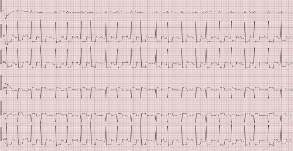

Importantly:
-
The rhythm was not atrial fibrillation, which is common in large-breed dogs.
-
P waves were present before every QRS complex.
-
A diagnosis of atrial tachycardia was made.
What Is Atrial Tachycardia?
The heart normally beats under control of a natural pacemaker. In atrial tachycardia, another area in the upper chambers of the heart generates rapid electrical signals, causing the heart to beat too fast.
When the heart beats too quickly:
-
There is less time for the heart to fill with blood between beats.
-
Blood output to the body decreases.
-
Over time, sustained fast rhythms can weaken heart muscle function.
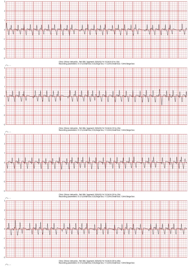
To eliminate the possibility of stress affecting the examination, we used CardioBird Vital Monitoring to continuously track the dog’s heart rhythm while discussing findings with the owners. We were more confident that the increased heart rate was pathological rather than stress-driven.
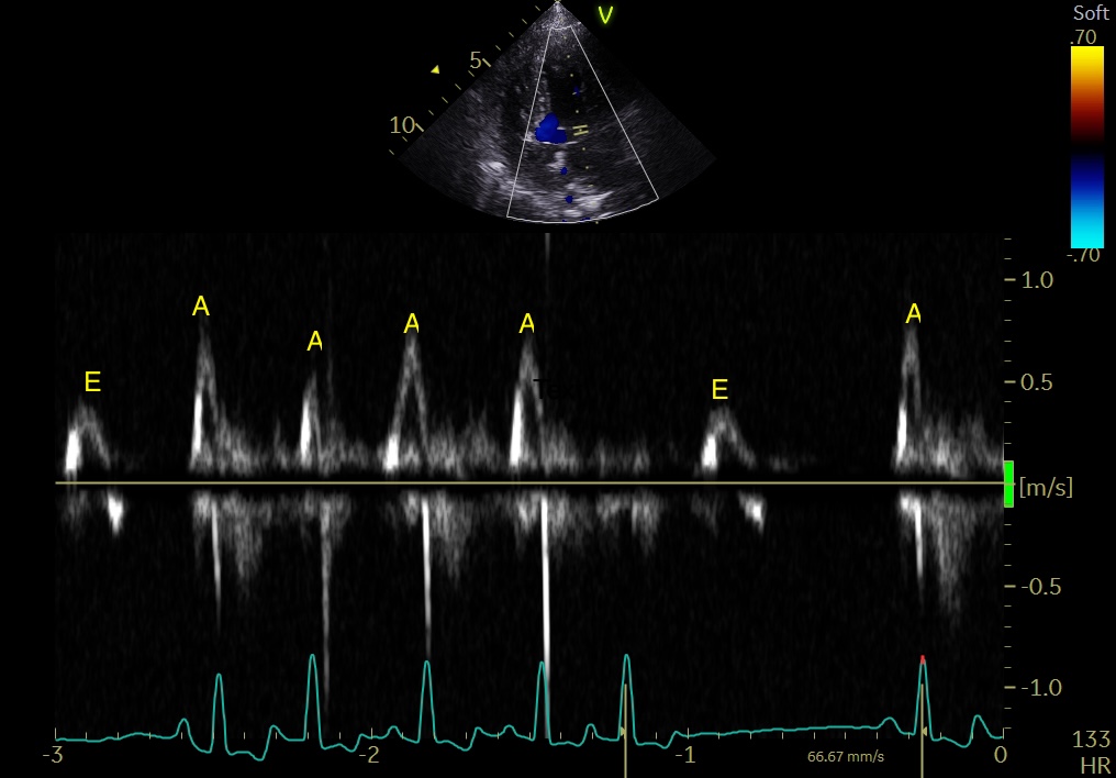
Echocardiography
Echocardiography showed reduced contraction and dyskinesia (uncontrolled movement) of the left ventricle. However, there was no evidence of dilated cardiomyopathy, a common heart muscle disease affecting large-breed dogs.
The reduced heart function was believed to be related to the persistent fast and irregular rhythm.
Possible Underlying Causes
Atrial tachycardia may be secondary to another condition rather than being a primary heart disease. Possible contributing factors include:
-
Neoplasia
-
Systemic Inflammation
-
Hyperthyroidism
-
Chronic pain or stress
-
Electrolyte imbalance
